R2OBBIE-3D


R2OBBIE-3D


Back to the
‘Physics of biology’ page
Back to the
“LANE”
introduction page

PUBLICATIONS
‣PLOS ONE (2015)

!! CHECK ALSO !!
Skin Cracking in Crocodiles
Science (2013)

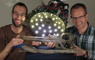
R2OBBIE-3D, a fast robotic high-resolution system
for quantitative phenotyping of surface geometry and colour-texture
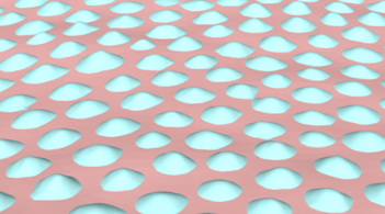
While recent imaging techniques provide insights into biological processes from the molecular to the cellular scale, phenotypes at larger scales remain poorly amenable to quantitative analyses. For example, investigations of the biophysical mechanisms generating skin morphological complexity and diversity would greatly benefit from 3D geometry and colour texture reconstructions. Here, we report on R2OBBIE-3D, an integrated system that combines a robotic arm, a high-resolution digital colour camera, an illumination basket of high-intensity light-emitting diodes and state-of-the-art 3D-reconstruction approaches. We demonstrate that R2OBBIE-3D is able to produce fully-textured 3D models of objects (including animals under anaesthesia), capturing both colour and geometry details from the scale of the meter down to the scale of about 11-25 microns (i.e., spanning five orders of magnitude) without the use of magnifying lenses. Moreover, the automation of R2OBBIE-3D makes the system highly versatile and allows for both practical scanning times and high-throughput analyses. R2OBBIE has the potential to greatly improve quantitative analyses of phenotypes in addition to providing multiple new applications in, e.g., biomedical science.
Supporting Material
The Supporting PDF file (‘Martins_etal_R2OBBIE_SupInfo_S1.pdf’) includes nine Supporting Figures and their legends, as well as links to 3D models used for the Supporting movies.
Supporting Movies
All movies are available on the PLOS ONE website as well as on the ‘EpiPhysX’ YouTube channel: https://www.youtube.com/channel/UCQwkb9mFahbopvMAVfJoPcA
✓Movie S1: http://youtu.be/EOBD8reW4dw
Supporting Movie S1 - Scanning with R2OBBIE-3D: the acquisition process. The movie shows the scanning process in Structure-from-Motion (PMVS) and Shape-from-Shading (PS) modes. See main text for details.
✓Movie S2: http://youtu.be/6WN-awvWZrM
Supporting Movie S2 - Corn Snake (Pantherophis guttatus): Geometry Reconstructed with Structure from Motion. The movie shows the surface geometry and colour texture of a scanned corn snake.
✓Movie S3: http://youtu.be/2lTWWJCgsmY
Supporting Movie S3 - Skeleton of a Sea Urchin: (Echinometra mathaei) Geometry Reconstructed with Photometric Stereo. The movie shows the surface geometry and colour texture of a scanned sea urchin skeleton.
✓Movie S4: http://youtu.be/p67wu5ZQSpA
Supporting Movie S4 - Coin of 0.5 Swiss Francs: Geometry Reconstructed with Photometric Stereo. The movie shows the surface geometry of the smallest Swiss coin.
✓Movie S5: http://youtu.be/sOBmVS15l8g
Supporting Movie S5 - Fossil of Marine Crocodylian: Geometry Reconstructed with Structure from Motion. The movie shows the surface geometry and colour texture of a 155-161 million-year-old fossil (head of a Metriorhynchus superciliosus, Upper Jurassic of England). Specimen PIMUZ A/III 14 from the Paläontologisches Institut und Museum, Universität Zürich, Switzerland.
Supporting Model Files
These correspond to the 3D models (2.4 to 14.7 millions faces) illustrated in Supporting Movies S2, S3, S4, and S5. After unzipping, each folder contains an ‘.obj’ file that can be opened and manipulated (zooming, panning, rotating) in any 3D viewer application supporting that file format, such as the free open-source software Meshlab [Cignoni P, Callieri M, Corsini M, et al. (2008) MeshLab: an Open-Source Mesh Processing Tool. Sixth Eurographics Italian Chapter Conference, 129-136.] available at http://meshlab.sourceforge.net. Note that the material file (‘.mtl’) and texturing files (‘.jpg’ or ‘.png’) must be kept in the same folder as the ‘.obj’ file when opening the latter. The Supporting_Model_S3_coin.zip file contains only an ‘.obj’ file as it is untextured. The largest 3D models should be opened on a reasonably recent computer equipped with a good graphical card and at least 4GB of RAM.
Be patient for download, these are big files (note the size of the file below) !
✓Supporting_Model_S1_snake.zip - 210 MB - (illustrated in Movie S2)
✓Supporting_Model_S2_urchin.zip - 365 MB - (illustrated in Movie S3)
✓Supporting_Model_S3_coin.zip - 277 MB - (illustrated in Movie S4)
✓Supporting_Model_S4_fossil.zip - 411 MB - (illustrated in Movie S5)
Publication
✓Martins A., Bessant M., Manukyan L. & M.C. Milinkovitch.
R2OBBIE-3D, a Fast Robotic High-Resolution System for Quantitative Phenotyping of Surface Geometry and Colour-Texture
PLOS ONE 10(6): e0126740 (2015) - doi:10.1371/journal.pone.0126740
![]() Supporting File 1 (Supp. Figs A-I, Legends of Supp. Movies S1-S5, Links to Supp. 3D-Model Files).
Supporting File 1 (Supp. Figs A-I, Legends of Supp. Movies S1-S5, Links to Supp. 3D-Model Files).
Coverage
Other related publications
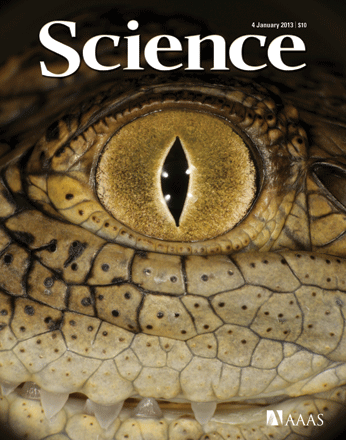
✓Milinkovitch M.C., Manukyan L., Debry A., Di-Poï N., Martin S., Dhillon D.S., Lambert D., Zwicker M.
Crocodile Head Scales Are Not Developmental Units But Emerge from Physical Cracking
Science 339, 78-81 (2013)
DOI: 10.1126/science.1226265
 Watch the Supplementary Movie (also available HERE and in YouTube)
Coverage
 Check the Movie illustrating the Computer Graphics Tools (version 2)
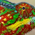
✓Teyssier J., Saenko S.V., van der Marel D. & M.C. Milinkovitch.
Photonic Crystals Cause Active Colour Change in Chameleons
Nature Communications 6: 6368 (2015)
![]() Supporting Information (Suppl Figs 1-4, Suppl Table 1, Suppl Text and Refs)
Supporting Information (Suppl Figs 1-4, Suppl Table 1, Suppl Text and Refs)
Coverage